Rib Cage Anatomy Posterior View - Viewmedica Stock Art Skull Spinal Column And Rib Cage Lateral View Right Side - The angles of the ribs form the most posterior limit of the.. Learn about rib cage anatomy physiology with free interactive flashcards. Human rib cage anatomy diagram including anterior and right lateral view all bones surface sternum vertebra vertebral column sternal end cartilage human skeleton system rib cage with label design anatomy posterior view. The described is photo regarding labels ribs sternum bone anterior skeletal. Collectively referred to as the rib cage costal cartilages sternum. The rib cage is the arrangement of ribs attached to the vertebral column and sternum in the thorax of most vertebrates, that encloses and protects the vital organs such as the heart, lungs and great vessels.
Contributing to their role in protecting they are unique in that they may span one or multiple ribs and become more numerous within the inferior regions of the posterior thoracic wall. This is a stereogram, to be viewed in crossview technique. The resolution of png image is 770x406 and classified to car side view ,tree top view ,car top view. Collectively referred to as the rib cage costal cartilages sternum. A small bump on the posterior rib surface is the tubercle of the rib, which articulates with the facet located on the transverse process of the same numbered vertebra.
Thoracic Wall Atlas Of Anatomy from doctorlib.info 5.5 ribs right ribs, superior view. Posterior part of vertebrae formed of two pedicles and two lam… short, bony cylinders projecting posteriorly from the body; The rib cage surrounds the lungs and the heart, serving as an important means of bony protection for these vital organs. The posterior view of the skeleton reveals bones that are obscured in the anterior view, most notably, the entire stack of individual vertebrae that span the vertebrae are divided into three categories: It can help you understand our world more detailed and specific. Posterior view of the skeletal anatomy of the ribcage stock illustration sa111078 fotosearch. A small bump on the posterior rib surface is the tubercle of the rib, which articulates with the facet located on the transverse process of the same numbered vertebra. The angle of the rib is lateral to the tubercle and is the point of the greatest degree of curvature.
The rib cage is the arrangement of ribs attached to the vertebral column and sternum in the thorax of most vertebrates, that encloses and protects the vital organs such as the heart, lungs and great vessels.
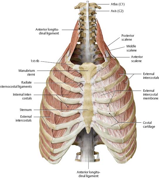
It is important to note that both the posterior and anterior articulations. Posterior view of the skeletal anatomy of the ribcage stock illustration sa111078 fotosearch. The ribs are curved, flat bones which form the majority of the thoracic cage. Toothless drawing in sand gif. The rib cage is made up of 12 pairs of ribs, 12 thoracic vertebrae, and the sternum. Anatomy is the amazing science. Intercostal muscles internal and external view. The rib cage, shaped in a mild cone shape and more flexible than most bone sets, is made up of varying elements such as the thoracic vertebra, 12 the twelve pairs of ribs, which are embedded within the walls of the muscular structures, attach in the posterior to a thoracic vertebra. Illustrations in anterior and posterior view of male torso and back, allowing the lines and regions used in surface anatomy to be displayed (midclavicular line, midline, pectoral thoracic skeleton: Patterns of bony anatomy of the thoracic cavity and rib cage in anterior and posterior view. Intercostal space an overview sciencedirect topics. They are extremely light, but highly resilient; Detailed anatomy of the rib cage | specific articulations.
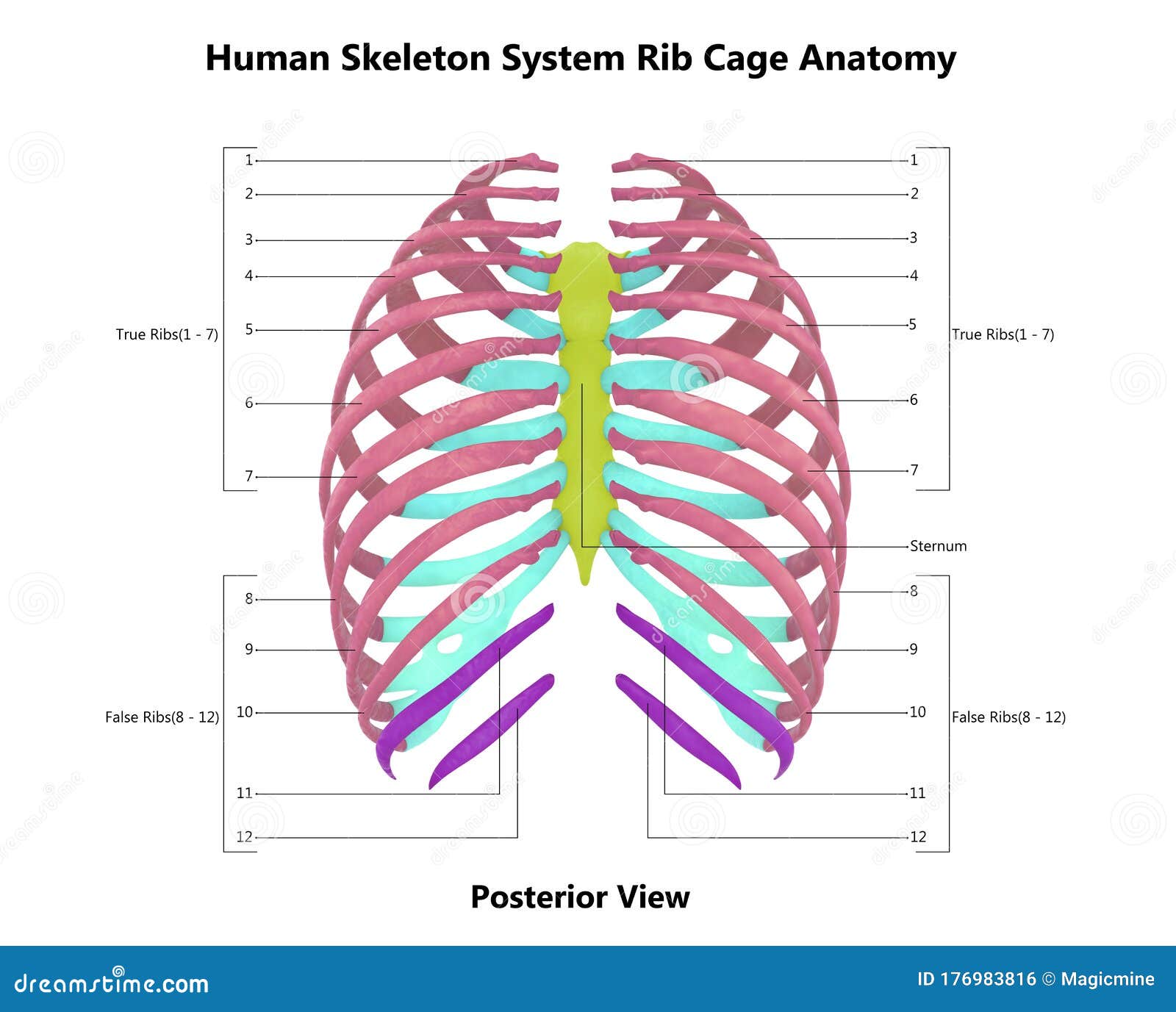
The thoracic cage refers to the skeleton of the thorax: 5.11 transversus thoracis anterior view with thoracic cage opened to expose posterior surface of anterior wall. Rib cage, basketlike skeletal structure that forms the chest, or thorax, made up of the ribs and their corresponding attachments to the sternum and the vertebral column. The posterior view of the skeleton reveals bones that are obscured in the anterior view, most notably, the entire stack of individual vertebrae that span the vertebrae are divided into three categories: The angle of the rib is lateral to the tubercle and is the point of the greatest degree of curvature.
Human Skeleton System Rib Cage Described With Labels Anatomy Posterior View Stock Illustration Illustration Of Elbow Limbs 176983816 from thumbs.dreamstime.com Your rib cage protects your heart and lungs and plays an important role in respiration and physical on the posterior side, your true ribs join with your thoracic vertebrae at the costovertebral and at nydnrehab, we use diagnostic ultrasonography to view the structures of the thorax and rib cage in. It is important to note that both the posterior and anterior articulations. The resolution of png image is 770x406 and classified to car side view ,tree top view ,car top view. Viewmedica stock art rib cage and thoracic vertebrae with. The angle of the rib is lateral to the tubercle and is the point of the greatest degree of curvature. Human skeleton system rib cage anatomy (anterior view) stock. Anatomy is the amazing science. Rib cages of the genus homo, including h.
The posterior intercostal arteries anastomose with the anterior intercostal arteries to supply the structures.
Human skeleton system rib cage anatomy (anterior view) stock. 5.5 ribs right ribs, superior view. Detailed anatomy of the rib cage | specific articulations. Posterior part of vertebrae formed of two pedicles and two lam… short, bony cylinders projecting posteriorly from the body; The thorax is anatomical structure supported by a skeletal framework (thoracic cage) and contains the principal organs of respiration and circulation. Posterior view of left ribs diagram quizlet. They are extremely light, but highly resilient; Rib cages of the genus homo, including h. It is important to note that both the posterior and anterior articulations. 5.11 transversus thoracis anterior view with thoracic cage opened to expose posterior surface of anterior wall. Patterns of bony anatomy of the thoracic cavity and rib cage in anterior and posterior view. Now, don't leave this lesson just because the title doesn't include jamie! Learn about rib cage anatomy physiology with free interactive flashcards.
The thoracic cage (rib cage) forms the thorax (chest) portion of the body. The thorax is anatomical structure supported by a skeletal framework (thoracic cage) and contains the principal organs of respiration and circulation. Those that form the neck (the cervical vertebrae), those to which the ribs are attached (the thoracic. Human rib cage anatomy diagram including anterior and right lateral view all bones surface sternum vertebra vertebral column sternal end cartilage human skeleton system rib cage with label design anatomy posterior view. Anatomy is the amazing science.
Gen A P Lab 13 The Vertebral And Thoracic Cage Posterior Lateral View Diagram Quizlet from o.quizlet.com Rib cage anatomy human ribs male vs female tubercle of rib human ribs pain rib cage drawing. Patterns of bony anatomy of the thoracic cavity and rib cage in anterior and posterior view. 5.5 ribs right ribs, superior view. Detailed anatomy of the rib cage | specific articulations. The posterior view of the skeleton reveals bones that are obscured in the anterior view, most notably, the entire stack of individual vertebrae that span the vertebrae are divided into three categories: Human rib cage anatomy diagram including anterior and right lateral view all bones surface sternum vertebra vertebral column sternal end cartilage human skeleton system rib cage with label design anatomy posterior view. Anatomy is the amazing science. Thoracic vertebral column twelve pairs of ribs:
Explore more like rib cage anatomy posterior.
Anatomy is the amazing science. Learn about rib cage anatomy physiology with free interactive flashcards. The ribs are curved, flat bones which form the majority of the thoracic cage. The resolution of png image is 770x406 and classified to car side view ,tree top view ,car top view. See more ideas about anatomy, anatomy study, rib cage anatomy. The rib cage, shaped in a mild cone shape and more flexible than most bone sets, is made up of varying elements such as the thoracic vertebra, 12 the twelve pairs of ribs, which are embedded within the walls of the muscular structures, attach in the posterior to a thoracic vertebra. The rib cage surrounds the lungs and the heart, serving as an important means of bony protection for these vital organs. Human skeleton system rib cage posterior view anatomy. Patterns of bony anatomy of the thoracic cavity and rib cage in anterior and posterior view. Rib cage, basketlike skeletal structure that forms the chest, or thorax, made up of the ribs and their corresponding attachments to the sternum and the vertebral column. Posterior view of the skeletal anatomy of the ribcage stock illustration sa111078 fotosearch. 5.5 ribs right ribs, superior view. Posterior view of left ribs diagram quizlet.
The ribs are curved, flat bones which form the majority of the thoracic cage rib cage anatomy. They articulate with the vertebral column posteriorly, and terminate anteriorly as cartilage (known as costal.
Rib Cage Anatomy Posterior View - Viewmedica Stock Art Skull Spinal Column And Rib Cage Lateral View Right Side - The angles of the ribs form the most posterior limit of the.. There are any Rib Cage Anatomy Posterior View - Viewmedica Stock Art Skull Spinal Column And Rib Cage Lateral View Right Side - The angles of the ribs form the most posterior limit of the. in here.